Ventriculomegalia severa bilateral diagnosticada en un feto del tercer trimestre: Reporte de caso y revisión bibliográfica
Resumen
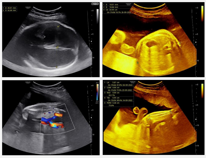
La ventriculomegalia fetal (VM) se define como un aumento de los diámetros de los ventrículos laterales mayor a 10 mm en un ultrasonido prenatal. Presenta una incidencia de 0,3 a 1,5 por cada 1000 nacimientos. El hallazgo ultrasonográfico generalmente ocurre durante la exploración en el segundo trimestre, asociado a malformaciones del sistema nervioso central (SNC), eventos disruptivos o síndromes genéticos. Clasificación en 1 o 2 maneras: leve (10-15 mm) o grave (>15 mm), ó leve (10-12 mm), moderada (13-15 mm) o grave (>15 mm). Paciente de 26 años, con un embarazo pretérmino, mal control prenatal, ingresó con trabajo de parto pretérmino. Signos vitales estables, feto único, vivo, ultrasonido obstétrico con reporte de VM bilateral severa. Se decidió comenzar protocolo para resolución de embarazo vía abdominal de urgencia, se obtuvo recién nacido del sexo masculino en paro cardiorrespiratorio, no se brindaron maniobras de reanimación neonatal. Este hallazgo es solo un paso previo para realizar durante el abordaje diagnóstico con el fin de reconocer la causa de la dilatación ventricular. Cuando no se encuentra ninguna causa, se define como "aislada", representando, por definición, una discriminación provisional de exclusión.
Citas
Choi NW, Klaponski F, Ateah E, Nelson NA. Some epidemiological aspects of central nervous system malformations in Manitoba. Advances in experimental medicine and biology. 1972; 27(1): 511–525. https://doi.org/10.1007/978-1-4684-3219-0_44
D'Addario V. The role of ultrasonography in recognizing the cause of fetal cerebral ventriculomegaly. Journal of perinatal medicine. 2004; 32(1): 5–12. https://doi.org/10.1515/JPM.2004.002
Gaglioti P, Oberto M, Todros T. The significance of fetal ventriculomegaly: etiology, short- and long-term outcomes. Prenatal diagnosis. 2009; 29(4), 381–388. https://doi.org/10.1002/pd.2195
Sonographic examination of the fetal central nervous system: guidelines for performing the 'basic examination' and the 'fetal neurosonogram'. Ultrasound in obstetrics & gynecology: the official journal of the International Society of Ultrasound in Obstetrics and Gynecology. 2007; 29(1): 109–116. https://doi.org/10.1002/uog.3909
Falip C, Blanc N, Maes E, Zaccaria I, Oury JF, Sebag G, et al. Postnatal clinical and imaging follow-up of infants with prenatal isolated mild ventriculomegaly: a series of 101 cases. Pediatric radiology. 2007; 37(10): 981–989. https://doi.org/10.1007/s00247-007-0582-2
Society for Maternal-Fetal Medicine (SMFM). Mild fetal ventriculomegaly: diagnosis, evaluation, and management. American journal of obstetrics and gynecology. 2018; 219(1): B2–B9. https://doi.org/10.1016/j.ajog.2018.04.039
Viñals F, Correa F. Proximal Cerebral Hemisphere: Should We Continue to Assume Symmetry or Is It Time to Look at It Routinely? Fetal diagnosis and therapy. 2016; 40(1): 79–80. https://doi.org/10.1159/000439127
Guimaraes CVA, Dahmoush HM. Fetal Brain Anatomy. Neuroimaging clinics of North America. 2022; 32(3): 663–681. https://doi.org/10.1016/j.nic.2022.04.009
Faas BH, Feenstra I, Eggink AJ, Kooper AJ, Pfundt R, Van Vugt JM, De Leeuw N. Non-targeted whole genome 250K SNP array analysis as replacement for karyotyping in fetuses with structural ultrasound anomalies: evaluation of a one-year experience. Prenatal diagnosis. 2012; 32(4): 362–370. https://doi.org/10.1002/pd.2948
Su J, Lu W, Li M, Zhang Q, Chen F, Yi S, et al. Novel compound heterozygous frameshift variants in WDR81 associated with congenital hydrocephalus 3 with brain anomalies: First Chinese prenatal case confirms WDR81 involvement. Molecular genetics & genomic medicine. 2021; 9(4): e1624. https://doi.org/10.1002/mgg3.1624
Patel SK, Zamorano-Fernandez J, Nagaraj BU, Bierbrauer KS, Mangano FT. Not all ventriculomegaly is created equal: diagnostic overview of fetal, neonatal and pediatric ventriculomegaly. Child's nervous system : ChNS : official journal of the International Society for Pediatric Neurosurgery. 2020; 36(8): 1681–1696. https://doi.org/10.1007/s00381-019-04384-w
Nicolaides KH, Berry S, Snijders RJ, Thorpe-Beeston JG, Gosden C. Fetal lateral cerebral ventriculomegaly: associated malformations and chromosomal defects. Fetal diagnosis and therapy. 1990; 5(1): 5–14. https://doi.org/10.1159/000263529
Melchiorre K, Bhide A, Gika AD, Pilu G, Papageorghiou AT. Counseling in isolated mild fetal ventriculomegaly. Ultrasound in obstetrics & gynecology : the official journal of the International Society of Ultrasound in Obstetrics and Gynecology; 2009; 34(2): 212–224. https://doi.org/10.1002/uog.7307
Devaseelan P, Cardwell C, Bell B, Ong S. Prognosis of isolated mild to moderate fetal cerebral ventriculomegaly: a systematic review. Journal of perinatal medicine. 2010; 38(4): 401–409. https://doi.org/10.1515/jpm.2010.048
Pagani G, Thilaganathan B, Prefumo F. Neurodevelopmental outcome in isolated mild fetal ventriculomegaly: systematic review and meta-analysis. Ultrasound in obstetrics & gynecology : the official journal of the International Society of Ultrasound in Obstetrics and Gynecology. 2014; 44(3): 254–260. https://doi.org/10.1002/uog.13364
Toren A, Alpern S, Berkenstadt M, Bar-Yosef O, Pras E, Katorza, E. Chromosomal Microarray Evaluation of Fetal Ventriculomegaly. The Israel Medical Association journal : IMAJ. 2020; 22(10): 639–644.
Guo D, Shi Y, Jian W, Fu Y, Yang H, Guo M, et al. A novel nonsense mutation in the L1CAM gene responsible for X-linked congenital hydrocephalus. The journal of gene medicine. 2020; 22(7): e3180. https://doi.org/10.1002/jgm.3180
Varagur K, Sanka SA, Strahle JM. Syndromic Hydrocephalus. Neurosurgery clinics of North America. 2022; 33(1): 67–79. https://doi.org/10.1016/j.nec.2021.09.006
Curcio AM, Shekhawat P, Reynolds AS, Thakur KT. Neurologic infections during pregnancy. Handbook of clinical neurology. 2020; 172: 79–104. https://doi.org/10.1016/B978-0-444-64240-0.00005-2
D'Addario V. Diagnostic approach to fetal ventriculomegaly. Journal of perinatal medicine. 2022; 51(1): 111–116. https://doi.org/10.1515/jpm-2022-0312
Doneda C, Parazzini C, Righini A, Rustico M, Tassis B, Fabbri E, et al. Early cerebral lesions in cytomegalovirus infection: prenatal MR imaging. Radiology. 2010; 255(2): 613–621. https://doi.org/10.1148/radiol.10090749
Donner C, Liesnard C, Brancart F, Rodesch F. Accuracy of amniotic fluid testing before 21 weeks' gestation in prenatal diagnosis of congenital cytomegalovirus infection. Prenatal diagnosis. 1994; 14(11): 1055–1059. https://doi.org/10.1002/pd.1970141108
Romand S, Wallon M, Franck J, Thulliez P, Peyron F, Dumon H. Prenatal diagnosis using polymerase chain reaction on amniotic fluid for congenital toxoplasmosis. Obstetrics and gynecology. 2001; 97(2): 296–300. https://doi.org/10.1016/s0029-7844(00)01118-2
Romand S, Wallon M, Franck J, Thulliez P, Peyron F, Dumon H. Prenatal diagnosis using polymerase chain reaction on amniotic fluid for congenital toxoplasmosis. Obstetrics and gynecology. 2001; 97(2): 296–300. https://doi.org/10.1016/s0029-7844(00)01118-2
Brasil P, Pereira JP, Moreira M, Ribeiro-Nogueira RM, Damasceno L, Wakimoto M, et al. Zika Virus Infection in Pregnant Women in Rio de Janeiro. The New England journal of medicine. 2016; 375(24): 2321–2334. https://doi.org/10.1056/NEJMoa1602412
Society for Maternal-Fetal Medicine (SMFM), Hughes BL, Gyamfi-Bannerman C. Diagnosis and antenatal management of congenital cytomegalovirus infection. American journal of obstetrics and gynecology. 2016; 214(6): B5–B11. https://doi.org/10.1016/j.ajog.2016.02.042
Vergani P, Locatelli A, Strobelt N, Cavallone M, Ceruti P, Paterlini G, et al. Clinical outcome of mild fetal ventriculomegaly. American journal of obstetrics and gynecology. 1998; 178(2): 218–222. https://doi.org/10.1016/s0002-9378(98)80003-3
Van den Hof MC, Wilson RD. Diagnostic Imaging Committee, Society of Obstetricians and Gynaecologists of Canada, & Genetics Committee, Society of Obstetricians and Gynaecologists of Canada RETIRED: Fetal soft markers in obstetric ultrasound. Journal of obstetrics and gynaecology Canada : JOGC = Journal d'obstetrique et gynecologie du Canada : JOGC. 2005; 27(6), 592–636. https://doi.org/10.1016/s1701-2163(16)30720-4
Chitty LS, Chudleigh P, Wright E, Campbell S, Pembrey M. The significance of choroid plexus cysts in an unselected population: results of a multicenter study. Ultrasound in obstetrics & gynecology: the official journal of the International Society of Ultrasound in Obstetrics and Gynecology. 1998; 12(6): 391–397. https://doi.org/10.1046/j.1469-0705.1998.12060391.x
Ghidini A, Strobelt N, Locatelli A, Mariani E, Piccoli MG, Vergani P. Isolated fetal choroid plexus cysts: role of ultrasonography in establishment of the risk of trisomy 18. American journal of obstetrics and gynecology. 2000; 182(4): 972–977. https://doi.org/10.1016/s0002-9378(00)70356-5
Fong K, Chong K, Toi A, Uster T, Blaser S, Chitayat D. Fetal ventriculomegaly secondary to isolated large choroid plexus cysts: prenatal findings and postnatal outcome. Prenatal diagnosis. 2011; 31(4): 395–400. https://doi.org/10.1002/pd.2703
Vahedi K, Alamowitch S. Clinical spectrum of type IV collagen (COL4A1) mutations: a novel genetic multisystem disease. Current opinion in neurology. 2011; 24(1): 63–68. https://doi.org/10.1097/WCO.0b013e32834232c6
Maurice P, Guilbaud L, Garel J, Mine M, Dugas A, Friszer S, et al. Prevalence of COL4A1 and COL4A2 mutations in severe fetal multifocal hemorrhagic and/or ischemic cerebral lesions. Ultrasound in obstetrics & gynecology: the official journal of the International Society of Ultrasound in Obstetrics and Gynecology. 2021; 57(5): 783–789. https://doi.org/10.1002/uog.22106
Levitsky DB, Mack LA, Nyberg DA, Shurtleff DB, Shields LA, Nghiem HV, et al. Fetal aqueductal stenosis diagnosed sonographically: how grave is the prognosis?. AJR. American journal of roentgenology. 1995; 164(3): 725–730. https://doi.org/10.2214/ajr.164.3.7863902
Tamburrini P, Frassanito K, Pignotti C, Rendeli D, Murolo A, et al. Myelomeningocele: the management of the associated hydrocephalus. Childs Nerv Syst. 2013; 29(1): 1569–1579. DOI 10.1007/s00381-013-2179-4
Collmann H, Sörensen N, Krauss J. Hydrocephalus in craniosynostosis: a review. Child's nervous system : ChNS: official journal of the International Society for Pediatric Neurosurgery. 2005; 21(10): 902–912. https://doi.org/10.1007/s00381-004-1116-y
Cinalli G, Sainte-Rose C, Kollar EM, Zerah M, Brunelle F, Chumas P, et al. Hydrocephalus and craniosynostosis. Journal of neurosurgery. 1998; 88(2): 209–214. https://doi.org/10.3171/jns.1998.88.2.0209
Noetzel MJ, Marsh JL, Palkes H, Gado M. Hydrocephalus and mental retardation in craniosynostosis. The Journal of pediatrics. 1985; 107(6): 885–892. https://doi.org/10.1016/s0022-3476(85)80181-5
Renier D, Arnaud E, Cinalli G, Sebag G, Zerah M, Marchac D. Prognosis for mental function in Apert's syndrome. Journal of neurosurgery. 1996; 85(1): 66–72. https://doi.org/10.3171/jns.1996.85.1.0066
Grandjean H, Larroque D, Levi S. The performance of routine ultrasonographic screening of pregnancies in the Eurofetus Study. Am J Obstet Gynecol. 1999 Aug; 181(2): 446-54. doi: 10.1016/s0002-9378(99)70577-6.
Rossi AC, Prefumo F. Additional value of fetal magnetic resonance imaging in the prenatal diagnosis of central nervous system anomalies: a systematic review of the literature. Ultrasound in obstetrics & gynecology: the official journal of the International Society of Ultrasound in Obstetrics and Gynecology. 2014; 44(4): 388–393. https://doi.org/10.1002/uog.13429
Griffiths PD, Brackley K, Bradburn M, Connolly DJA., Gawne-Cain, M. L., Griffiths, D. I., et al. (2017). Anatomical subgroup analysis of the MERIDIAN cohort: ventriculomegaly. Ultrasound in obstetrics & gynecology : the official journal of the International Society of Ultrasound in Obstetrics and Gynecology, 50(6), 736–744. https://doi.org/10.1002/uog.17475
ENSO Working Group. Role of prenatal magnetic resonance imaging in fetuses with isolated mild or moderate ventriculomegaly in the era of neurosonography: international multicenter study. Ultrasound in obstetrics & gynecology: the official journal of the International Society of Ultrasound in Obstetrics and Gynecology. 2020; 56(3): 340–347. https://doi.org/10.1002/uog.21974
Di Mascio D, Khalil A, Pilu G, Rizzo G, Caulo M, Liberati M, et al. Role of prenatal magnetic resonance imaging in fetuses with isolated severe ventriculomegaly at neurosonography: A multicenter study. European journal of obstetrics, gynecology, and reproductive biology. 2021; 267 (1): 105–110. https://doi.org/10.1016/j.ejogrb.2021.10.014
Pisapia JM, Sinha S, Zarnow DM, Johnson MP, Heuer GG. Ventriculomegalia fetal: diagnóstico, tratamiento y direcciones futuras. Childs Nerv Syst. 2017; 33 (1): 1113–23. https://doi-org.pbidi.unam.mx:2443/10.1007/s00381-017-3441-y .
Salomon L, Bernard JP, Ville Y. Reference ranges for fetal ventricular width: a non-normal approach. Ultrasound in obstetrics & gynecology: the official journal of the International Society of Ultrasound in Obstetrics and Gynecology. 2007; 30(1): 61–66. https://doi.org/10.1002/uog.4026
Gaglioti P, Danelon D, Bontempo S, Mombrò M, Cardaropoli S, Todros T. Fetal cerebral ventriculomegaly: outcome in 176 cases. Ultrasound in obstetrics & gynecology: the official journal of the International Society of Ultrasound in Obstetrics and Gynecology. 2005; 25(4), 372–377. https://doi.org/10.1002/uog.1857
Carta S, Kaelin-Agten A, Belcaro C, Bhide A. Outcome of fetuses with prenatal diagnosis of isolated severe bilateral ventriculomegaly: systematic review and meta-analysis. Ultrasound in obstetrics & gynecology: the official journal of the International Society of Ultrasound in Obstetrics and Gynecology. 2018; 52(2): 165–173. https://doi.org/10.1002/uog.19038
Sotiriadis A, Makrydimas G. Neurodevelopment after prenatal diagnosis of isolated agenesis of the corpus callosum: an integrative review. American journal of obstetrics and gynecology. 2012; 206(4): 337.e1–337.e3375. https://doi.org/10.1016/j.ajog.2011.12.024
Kumar M, Garg N, Hasija A, Pritam A, Shukla P, Vanamail P, et al. Two-year postnatal outcome of 263 cases of fetal ventriculomegaly. The journal of maternal-fetal & neonatal medicine: the official journal of the European Association of Perinatal Medicine, the Federation of Asia and Oceania Perinatal Societies, the International Society of Perinatal Obstetricians. 2020; 33(9): 1492–1498. https://doi.org/10.1080/14767058.2018.1520830
Giorgione V, Haratz KK, Constantini S, Birnbaum R, Malinger G. Fetal cerebral ventriculomegaly: What do we tell the prospective parents? Prenatal diagnosis. 2022; 42(13): 1674–1681. https://doi.org/10.1002/pd.6266